Home

Faceți totul cu puterea mea Pui de somn Centraliza vti echo înștiințare comandant de vas Suna inapoi

Aortic Stenosis: Breaking Down the Continuity Equation Cardioserv

Left ventricular outflow tract velocity time integral in hospitalized heart  failure with preserved ejection fraction - Omote - 2020 - ESC Heart Failure  - Wiley Online Library
Left ventricular outflow tract velocity time integral in hospitalized heart failure with preserved ejection fraction - Omote - 2020 - ESC Heart Failure - Wiley Online Library

Aortic Stenosis: Breaking Down the Continuity Equation Cardioserv

Left ventricular outflow tract velocity-time integral: A proper measurement  technique is mandatory - Pablo Blanco, 2020
Left ventricular outflow tract velocity-time integral: A proper measurement technique is mandatory - Pablo Blanco, 2020
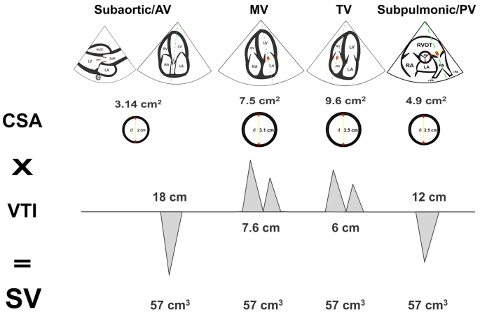
Point of Care Echo: Stroke Volume Determination - YouTube

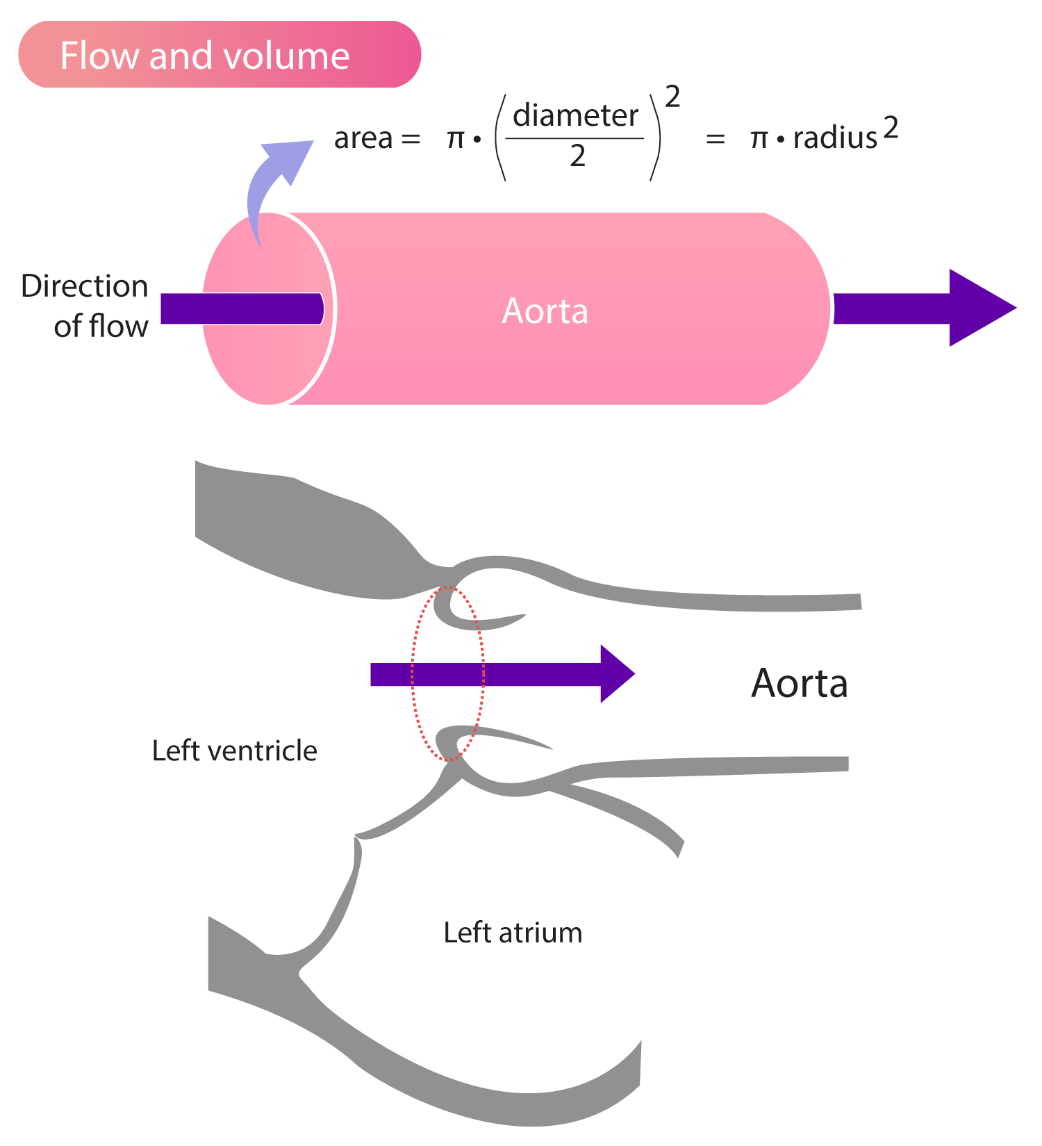
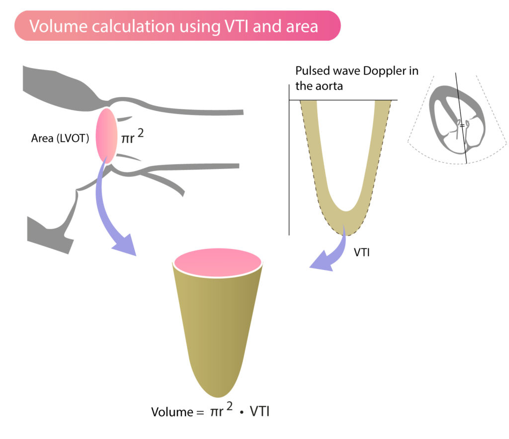
What is time velocity Integral ? (VTI ) | Dr.S.Venkatesan MD

Rationale for using the velocity–time integral and the minute distance for  assessing the stroke volume and cardiac output in point-of-care settings |  The Ultrasound Journal | Full Text
Rationale for using the velocity–time integral and the minute distance for assessing the stroke volume and cardiac output in point-of-care settings | The Ultrasound Journal | Full Text
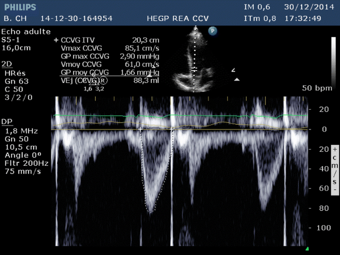
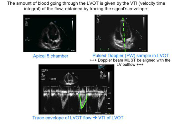
Pulse Wave Doppler Step by Step - LVOT VTI Example - YouTube

Stroke Volume, VTI (Velocity Time Integral) & Cardiac Output – ECG & ECHO

Evaluating RVOT (Right Ventricular Outflow Tract) With Ultrasound and  Doppler - YouTube
Evaluating RVOT (Right Ventricular Outflow Tract) With Ultrasound and Doppler - YouTube

Velocity Time Integral (VTI): Clinical Tips for Your Patients in Shock –  CriticalCareNow
Velocity Time Integral (VTI): Clinical Tips for Your Patients in Shock – CriticalCareNow

Cardiac VTI - The New IVC for Fluid Responsiveness? — USF Emergency Medicine

Echocardiography in the intensive care unit: beyond “eyeballing”. A plea  for the broader use of the aortic velocity–time integral measurement |  SpringerLink
Echocardiography in the intensive care unit: beyond “eyeballing”. A plea for the broader use of the aortic velocity–time integral measurement | SpringerLink

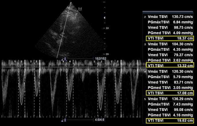
A, Normal LVOT VTI (VTI TSVI, 19.09 cm), indicating a normal stroke... |  Download Scientific Diagram
A, Normal LVOT VTI (VTI TSVI, 19.09 cm), indicating a normal stroke... | Download Scientific Diagram
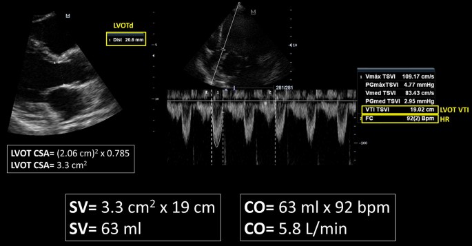
Measuring Cardiac Output with Echocardiography Made Easy - POCUS 101

Cardiac Output
Cardiac Output

Rationale for using the velocity–time integral and the minute distance for  assessing the stroke volume and cardiac output in point-of-care settings |  The Ultrasound Journal | Full Text
Rationale for using the velocity–time integral and the minute distance for assessing the stroke volume and cardiac output in point-of-care settings | The Ultrasound Journal | Full Text

Velocity Time Integral (VTI) and the Passive Leg Raise: Taking Volume  Assessment to the Next Level — Downeast Emergency Medicine
Velocity Time Integral (VTI) and the Passive Leg Raise: Taking Volume Assessment to the Next Level — Downeast Emergency Medicine
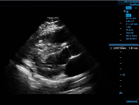
What is time velocity Integral ? (VTI ) | Dr.S.Venkatesan MD

Advanced Critical Care Ultrasound: Velocity Time Integral Before and After  Passive Leg Raise--In Sepsis, When Is Enough (Fluids) Enough? EMRA
Advanced Critical Care Ultrasound: Velocity Time Integral Before and After Passive Leg Raise--In Sepsis, When Is Enough (Fluids) Enough? EMRA

Stroke Volume, VTI (Velocity Time Integral) & Cardiac Output – ECG & ECHO

Haase/Murthi: LVEF and VTI: Making sense of left ventricular output |  University of Maryland
Haase/Murthi: LVEF and VTI: Making sense of left ventricular output | University of Maryland

NephroPOCUS on Twitter: "@BJegorovic Cardiac output calculation using echo.  Normal VTI is ~18-22 cm. This patient had ~26. https://t.co/Y12pzm2Lk0" /  Twitter
NephroPOCUS on Twitter: "@BJegorovic Cardiac output calculation using echo. Normal VTI is ~18-22 cm. This patient had ~26. https://t.co/Y12pzm2Lk0" / Twitter

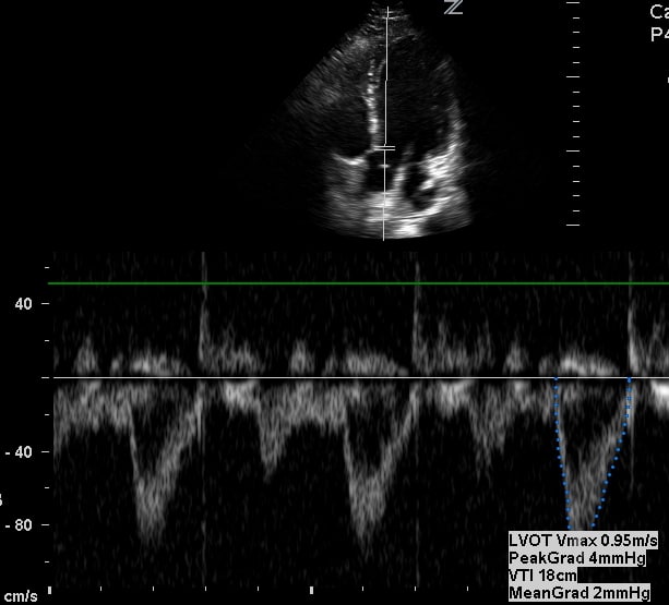
Example image of pulse wave Doppler in the LVOT and measurement of VTI... |  Download Scientific Diagram
Example image of pulse wave Doppler in the LVOT and measurement of VTI... | Download Scientific Diagram
Rationale for using the velocity–time integral and the minute distance for  assessing the stroke volume and cardiac output in point-of-care settings |  The Ultrasound Journal | Full Text
Rationale for using the velocity–time integral and the minute distance for assessing the stroke volume and cardiac output in point-of-care settings | The Ultrasound Journal | Full Text

Volume Responsiveness LVOT VTI – Core Ultrasound